Subdural Or Subarachnoid Hemorrhage
The dura mater and arachnoid are two of the three meninges that humans have. The meninges are the structures that line the central nervous system. A subdural or subarachnoid hemorrhage refers to a hemorrhage that occurs below each of these two meninges.
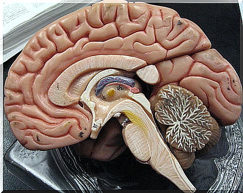
The meninges
The brain and spinal cord are protected by the skull and spinal column, but they also have another protection system: the meninges, also involved in nervous development.
As the NIH Cancer Dictionary explains: “The meninges are the three layers of tissue that cover and protect the brain and spinal cord ”. We have three of these layers, which from the outside to the inside are:
- Dura mater.
- Arachnoid.
- Pia mater.
The outermost and thickest, the dura, is separated from the bone by the epidural space. In the skull, this space is virtual, that is, it does not exist because the dura is attached to the bone. However, it does exist in the spinal cord and is occupied by veins and fat.
Below the dura is the arachnoid, separated by the subdural space. This space is also virtual and only becomes real when a hemorrhage occurs and the blood separates the two meninges.
The arachnoid sends a series of extensions that run through the subarachnoid space towards the pia mater. The subarachnoid space is occupied by cerebrospinal fluid that is responsible, among other things, for damping pressure changes due to blows or sudden movements.
Finally, the pia mater is intimately attached to the nervous tissue, accompanying it even in its grooves. The possibility of even accompanying him to the interior of the tissue is under investigation.
Subdural or subarachnoid hemorrhage
In a subdural or subarachnoid hemorrhage, the first circumstance is the exit of blood from the blood vessels, which is stored in the spaces between the meninges. This causes damage to the brain tissue, thus generating various clinical pictures.
However, depending on whether the hemorrhage is subdural or subarachnoid, the triggers, the course of the pathology and its symptoms will be different.
Subdural hemorrhage
Subdural hemorrhage is defined as the collection of blood in the virtual space between the dura mater and the arachnoid. This blood is usually of venous origin and usually responds to traumatic causes. However, there are three types of subdural hematoma depending on the time it takes to become apparent:
- Acute subdural hematoma.
- Subacute subdural hematoma.
- Chronic subdural hematoma.
Acute subdural hematoma
It is the one that is evidenced earlier. It is usually due to severe trauma that tears the veins from the cerebral cortex to the meninges.
Whoever experiences it usually falls immediately into a coma. In addition, signs of hemispheric focality usually appear. This means that a specific part of the brain stops working. Some examples of focality are:
- Hemiparesis: partial impotence for movement due to injury to the area that governs motor skills.
- Mydriasis: abnormal increase in pupillary diameter due to injury to the area that controls the iris muscle.
Subacute subdural hematoma
It evolves somewhat slower, and is usually less serious. This is because the amount of extravasated blood is less and clotting mechanisms can slow down the bleeding. Its cause is also usually traumatic in nature.
In the first place, consciousness is usually lost and then regained. Afterwards, for several days you will experience progressive clouding, in addition to signs of focality.
Chronic subdural hematoma
It is the result of multiple minor trauma over time. These give rise to small extravasations of blood that, when not reabsorbed, end up giving rise to a subdural hematoma of considerable dimensions. It is relatively common in the elderly.
The early symptom is usually headache or headache, associated with alterations in affectivity and behavior. The deterioration is progressive, with a tendency to sleep, slowed thinking and others.
Subarachnoid hemorrhage
Subarachnoid hemorrhage is defined as the collection of blood between the arachnoid and the pia mater. Blood is usually of arterial origin and has various causes. The most common is the rupture of an aneurysm , but it can also be due to vascular malformations.
Aneurysms can present with headaches or seizures before they rupture. In up to a third of the cases, the triggering factor for the rupture of the aneurysm is a physical effort with an emotional component or a long exposure to the sun.

Once they rupture, the subarachnoid hemorrhage begins. The most common is that it occurs between the ages of 40 and 60. The beginning of the clinic is abrupt and appear:
- Vomiting
- Clouding.
- Very severe headache.
- Photophobia (intolerance to light due to pain or discomfort).
Around 48 hours later, the meningeal syndrome usually appears, due to irritation of the meninges. Thus, the above symptoms are joined by a stiff neck. Focal deficits may also appear, such as paralysis of eye movements.
Subarachnoid hemorrhages cause sequelae in up to 60% of those who experience them. In addition, 40% of the survivors develop some type of dependency.