Human Chorionic Gonadotropin (hCG)
Human chorionic gonadotropin (hCG) is a hormone produced during the first weeks of pregnancy by the embryo. It is also produced by the cells of some types of tumor.
With regard to pregnancy, the concentration of this hormone, either in urine or blood, is what is measured with pregnancy tests . The tests commercial (which can be done at home) detected in the urine. In the hospital setting, it is usually analyzed in blood, as it is more accurate.
Structure of hCG
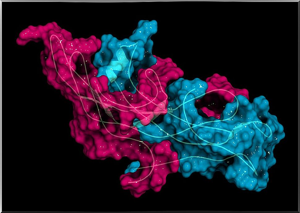
Each hCG molecule is made up of two subunits, called alpha and beta .
- The alpha subunit is common to other hormones, luteinizing hormone (LH) or follicle stimulating hormone (FSH).
- Both LH and FSH are hormones released into the blood from the pituitary gland with activity on the gonads.
- In contrast, the beta subunit is different and specific to hCG.
Where is it produced?
HCG is produced and released by syncytiotrophoblast cells . This is a group of cells, similar to a mass, that allow the attachment of the embryo to the placenta.
After fertilization, the first cell divisions of the future embryo take place. After 5 or 6 days, this “egg” is called a blastocyst . Two different cell groups will be generated from it:
- An internal one, called a trophoblast, which will give rise to the embryo.
- An external one, the syncytiotrophoblast , which allows the embryo to attach to the placenta.
B-hCG and pregnancy
The embryonic cells begin to produce the hormone towards the end of the first week of pregnancy. From this moment, production increases progressively until days 60 or 70, moment of maximum production. Thereafter, it gradually decreases until the levels stabilize and keep them constant until the end of the pregnancy.
The tests pregnancy measure the concentration of b -hCG in the urine, but can also be measured in maternal blood and in amniotic fluid. In this sense, the concept of discrimination zone of b -hCG is of special importance .
It is the concentration of hormone in the blood from which the gestational sac should be seen by ultrasound. It corresponds to 1500-2000 U / ml.
What functions does it have during pregnancy?

- It is believed to act on the mother’s immune system , inhibiting the lymphocyte response. Thus, they do not recognize the embryo as an antigen and do not trigger an immune response against it, allowing implantation.
- It is responsible for maintaining the corpus luteum until the placenta produces enough progesterone on its own. The corpus luteum is a follicular structure that produces hormones, including progesterone, which works by “maintaining” the pregnancy.
- Stimulates the synthesis of estrogens.
- It acts on the fetal testicle, stimulating the production of testosterone in it around week 10.
Ectopic pregnancy and multiple pregnancy
An ectopic pregnancy is one in which the fertilized egg implants at points other than the uterine wall. The most common sites are the fallopian tubes, the abdomen, or the pelvic cavity. Besides being an unviable pregnancy, it is a very high life risk for the mother.
When should an ectopic pregnancy be suspected?
- In cases of empty ultrasound and b-hCG concentrations that should allow the gestational sac to be visualized.
- When b-hCG values are lower than expected for the time of pregnancy.
In contrast, during multiple pregnancies the opposite happens: hCG levels skyrocket. In these cases, after the test, by means of an ultrasound the diagnosis of twin pregnancy can be established without problem.
B-hCG and tumors
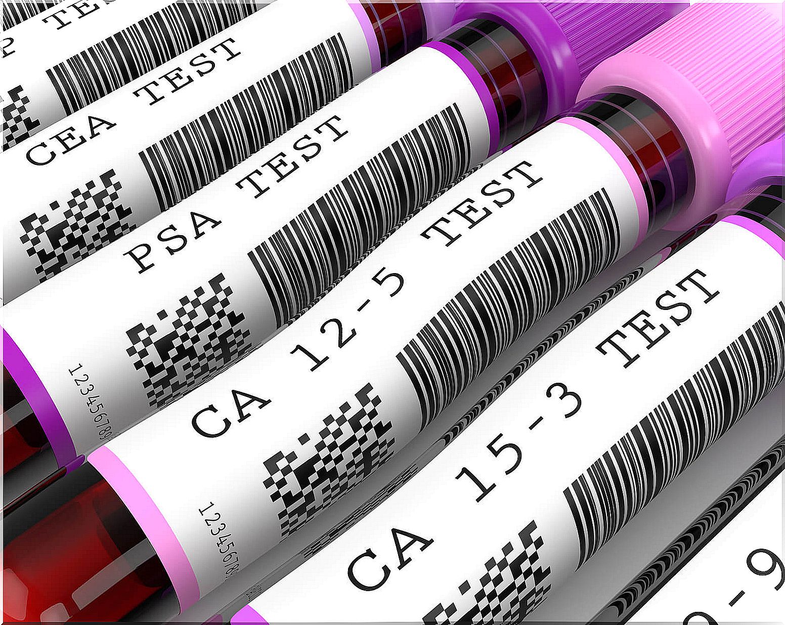
Human chorionic gonadotropin, in addition to being used in the detection and control of pregnancy, can be used as a tumor marker. This is because some placental, trophoblastic, and germ cell-derived tumors also produce this hormone. Some examples of tumors include choriocarcinoma, teratomas, and some seminomas.
Choriocarcinoma is a malignant and generally very aggressive tumor derived from trophoblastic cells (cells that help form the placenta). It metastasizes frequently, generally to the lungs.
Teratomas derive from the three cell sheets from which all structures in the embryo are formed. Due to this, they are tumors in which all kinds of components can be found, such as teeth, hair, eyes …
Some testicular germ cell tumors, called seminomas. They are rare tumors, generally affecting young men. In these cases, alpha-fetoprotein (AFP) and hCG levels rise.